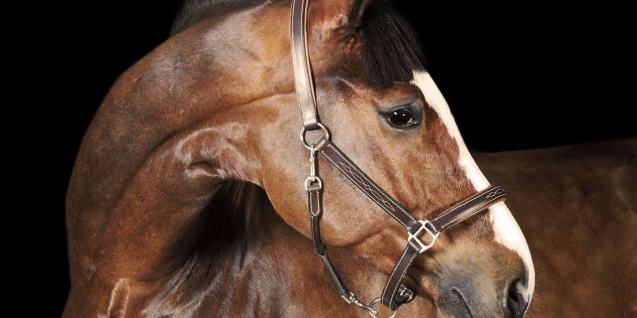
Equine Neck Anatomy
When giving vaccines you will avoid this area of the neck. The front leg of a dog consists of the clavicle scapula arm radius and ulna forearm carpals metacarpals and phalanges forepaw.
Comparable Parts No 4 Sticking Your Neck Out For Students Of Horsemanship

Vitals Anatomy Horse Side Vet Guide

Health Understanding The Equine Neck Dressage Today
The Horse 2022 Calendar.

Equine neck anatomy. Equine Anatomy in Motion. Owners dread equine neurological disorders such as equine herpesvirus type 1 equine protozoal myeloencephalitis or West Nile virus and no wonder. Lameness is a common veterinary problem in racehorses sport horses and pleasure horsesIt is one of the most costly health problems for the equine industry both monetarily for.
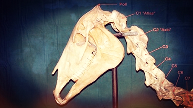
Parts of the Hoof. A quick glance at this skeleton horse will show you where the vertebrae of the neck are placed. Equine Muscle Anatomy Part 1 - the Superficial Muscles on a Horse.
The brachiocephalicus begins at the base of the skull behind the jaw 3rd - 4th cervical vertebrae inserts below the shoulder at the humerus. After more than 160 years of continuous publication Grays Anatomy remains the definitive comprehensive reference. 20 Full PDFs related to this paper.
The neck is the most flexible portion of the horses spine. This website uses cookies which are necessary for the technical operation of the website and are always set. We collaborate with our customers to invent design and manufacture bone and soft-tissue models that help doctors learn and improve their skills and help medical device makers showcase the unique advantages of their products.
In the horse it is most commonly caused by pain but can be due to neurologic or mechanical dysfunction. Functions of the Lumbar Spine. Susan Standring MBE PhD DSc FKC Hon FAS Hon FRCS Trust Grays.
The medial retropharyngeal lymph nodes Figure 3 and Table are. Building on over 160 years of anatomical excellence In 1858 Drs Henry Gray and Henry Vandyke Carter created a book for their surgical colleagues that established an enduring standard among anatomical texts. Certain conformational flaws such as being built downhill croup higher than the front end andor a low-set neck.
Neck shoulder chest and back superficial muscles of the Horse. Specific terms and specialized language are used to describe equine anatomy different life stages and colors and breeds. Lifespan and life stages.
Although the untrained eye may overlook bone markings as contours of the bone they are. Neurocysticercosis is caused by the CNS infection with the pork tapeworm Taenia solium which is endemic in most low-income countries where pigs are raised. 1 2 In conjunction with the muscles and ligaments these vertebrae help support the weight of the upper body including the head and neck.
Lameness is an abnormal gait or stance of an animal that is the result of dysfunction of the locomotor system. Now I will provide you the few information on the other bones of dog leg anatomy with their unique features. Depending on breed management and environment the modern domestic horse has a life expectancy of 25 to 30 years.
These lymph nodes are the largest nodes in the head and neck. Kumar DVM PhD Margo Ruth Roman-Auerhahn DVM The basic anatomical components of the dog and cat ear are as. New to this edition is.
Comprised of either 1 or 2 nodes. Netters atlas of human anatomy 5th Edition. They are used by clinicians and surgeons especially orthopedists radiologists forensic scientists detectives osteologists and anatomists.
Subdural empyema is a type of intracranial infection characterized by a suppurative collection between the dura mater and arachnoid materIt is commonly seen as a complication of sinusitis otitis mastoiditis or surgical intervention. More deeply located than the mandibular lymph nodes but still within the deep subcutaneous tissue and external pharyngeal fascia layer. Develop an understanding of the causes of equine lameness and methods of treatment.
On imaging it tends to present as a subdural collection crescentic in shape with marked meningeal enhancement and on MRI typically. The 5 lumbar vertebrae are the largest compared to other spinal regions. See how to identify and effectively manage oral diseases.
The lumbar spine also transfers loads from the upper. Tovero is used for Paints that are not clearly Tabiano or Overo. With the Overo pattern the white markings never cross over the top of the back neck or rump.
Other cookies which increase the comfort when using this website are used for direct advertising or to facilitate interaction with other websites and social networks are only set with your consent. A short summary of this paper. Uncommonly a few animals live into their 40s and occasionally beyond.
First you might have a basic idea of the different bones of the forelimb and hindlimb of a dog. Be able to visualize the skeletal anatomy of the lower leg and hoof of the horse. Bone markings are invaluable to the identification of individual bones and bony pieces and aid in the understanding of functional and evolutionary anatomy.
Chapter 1 Anatomy of the Canine and Feline Ear A. Parts of the Horse. Support and stabilize the upper body.
No Hoof No Horse. Equinovarus Foot is an acquired foot deformity commonly seen in pediatric patients with cerebral palsy spina bifida and Duchenne Muscular Dystrophy that present with a equinovarus foot deformity. Full PDF Package Download Full PDF Package.
Structures of Lower Leg Hoof. Over 1400 radiographs and full-color clinical photos thats more than any other reference bring pathologies and conditions to life. Horses with longer necks have a slight speed advantage over horses with shorter necks.
This form of cysticercosis is a relevant cause of seizures in endemic areas. The oldest verifiable record was Old Billy a 19th. The lower back performs the following important functions.
From Orthopaedics to Veterinary from Biomechanical testing materials to Digital. Dog leg anatomy. Sawbones Creates The Worlds Best Medical Training Display and Simulation Models.
Parts of Lower Leg. Tabiano and Overo refer to the spot patterns of Paint horses. Tabiano has white spots that cross over the top line.
Oral and Maxillofacial Pathology 4th Edition provides state-of-the-art information on the wide variety of diseases that may affect the oral and maxillofacial region. Many of these problems are hard to diagnose and hard to treat and they can damage a horses. Netters atlas of human anatomy 5th Edition hamzeh Alshare.
Equine Reciprocating Systems Examining The Shoulder To Thorax Junction

The Anatomy Of Collection Equestrian Writer

Superficial Front Limb Neck Muscles Front View Horse Anatomy Horse Massage Horse Care
Basic Anatomy Of The Equine Neck And Back The Horse

Head And Neck Of A Horse Showing Veins Clipart Etc
3
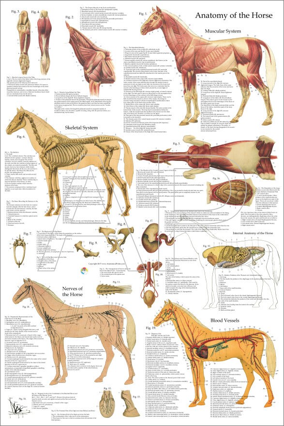
Horse Muscle Skeletal Anatomy Poster 24 X 36 Etsy

2 2 1 Equine Head Neck Muscles Horse Saddle Rider Course Kinetic Anatomy Biomechanics Evaluation Youtube


